MRI – Magnetic Resonance Imaging
Magnetic Resonance Imaging (MRI) provides clear, detailed images of internal organs and tissues without using X-ray radiation, making it valuable for diagnosing a wide range of conditions.
CT – Computed Tomography
Computed Tomography (CT) scans provide detailed, cross-sectional images of various tissues, making it easier to diagnose conditions like cancers, cardiovascular disease, and musculoskeletal disorders.
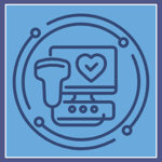
Ultrasound
Ultrasound imaging uses high-frequency sound waves to capture detailed images of internal organs and blood flow, providing a safe and effective way to examine areas like the heart, liver, and kidneys without using X-ray radiation.
Digital Mammography
Mammography uses low-dose X-rays for early breast cancer detection, with advanced full-field digital mammography and Computer-Aided Detection (CAD) to enhance accuracy and comfort for patients.
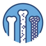
Bone Density Testing
Bone Density (DEXA) testing is essential for diagnosing osteoporosis and monitoring treatment, helping assess fracture risk and bone health, especially for postmenopausal women.
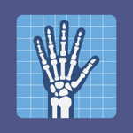
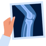
X-Ray – Bone Radiography
Mammography uses low-dose X-rays for early breast cancer detection, with advanced full-field digital mammography and Computer-Aided Detection (CAD) to enhance accuracy and comfort for patients.
Echocardiogram
An echocardiogram uses ultrasound to capture real-time images of the heart’s structure, helping diagnose conditions like valve disease and heart defects in a simple, non-invasive procedure.
Arthrogram
An arthrogram is a medical imaging test that uses contrast dye and X-ray, CT, or MRI to get detailed pictures of a joint.
About Us
Monument Imaging & Diagnostic Center has proudly served the Jacksonville community since 2000 as a trusted, free-standing, full-modality medical imaging center. Our commitment to patient care is matched by our use of state-of-the-art technology and the expertise of our on-site, Board-Certified radiologists, certified by the American Board of Radiology. With a diverse background in both hospital-based and outpatient imaging, our radiologists provide quality and precision in every diagnosis.
As an American College of Radiology (ACR) accredited facility, we uphold the highest practice standards, ensuring accuracy and peace of mind. We offer flexible, same-day or next-day appointments to accommodate your schedule, and your doctor will receive a detailed radiologist report within 24 to 48 hours.
Choose Monument Imaging & Diagnostic Center for prompt, reliable, and compassionate diagnostic care, providing assurance and clarity for you and your family.
Ready to take the next step? Let us help you bring clarity to your health issues. Call us for an appointment to have an imaging and diagnostic test taken.
Call us at (904) 855-0700
Testimonials
The staff was beyond professional from the front desk all the way through my appointment. I had a mammogram and ultrasound performed and was able to walk out of my appointment with the comfort that a doctor had review my images and the mass was not cancer. I highly recommend 9A Imaging to anyone who may need their services.
Amelia
I’ve always been treated beautifully there and I’ve been seen there twice… I’ve never had any issues with my insurance nor with billing…
Tracy
My husband had a MRI Arothrogram done here. The staff is friendly and helpful. The MRI itself was not bad at all. It went fairly quickly. They had to numb the area and then inject contrast into the joint area…..We would return to do imaging again here if needed.